Gastrointestinal Stromal Tumor (GIST)

A gastrointestinal stromal tumor (GIST) is a relatively rare type of cancer that most commonly develops in the stomach. However, these tumors can occur anywhere along the digestive tract — from the esophagus (swallowing tube) to the stomach, small intestine, colon and rectum.
GISTs grow in the muscle of the digestive tract wall and are derived from pacemaker cells, the cells that stimulate movement of the gut. They are classified as sarcomas — tumors of connective tissue — and are different from adenocarcinomas, which are more common in the digestive tract. The discovery of a molecular switch that turns on the growth of these tumors has made it possible to diagnose GISTs and target their growth with specific drugs.Because this cancer is uncommon, it’s important to find a care team that’s experienced in identifying, treating and researching GISTs. The University of Chicago Medicine GIST team is led by physicians with extensive experience and expertise with these tumors.
We also understand how a maze of tests, doctor’s visits and consultations can be overwhelming. Our medical and surgical oncologists, interventional endoscopists, pathologists and nurses work together to address each patient’s needs every step of the way. We’ll help you obtain all of the necessary tests you need as quickly and efficiently as possible.
You may not notice early GIST symptoms. Because these tumors are so rare, your doctor may not recognize them either. Without early detection and treatment, a GIST tumor can become large or spread (metastasize) to other parts of the body, such as the liver or abdominal cavity.
To help you receive a precise diagnosis, we offer several procedures:
Computed Tomography (CT scan)
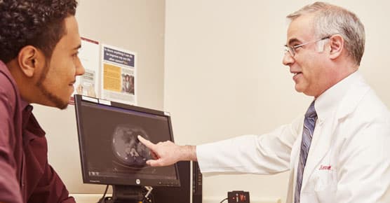
CT scans help identify the precise location of a tumor and determine if it has spread to other organs. UChicago Medicine offers the most sophisticated CT scans capable of creating vivid, three-dimensional images of GISTs.
Esophagogastroduodenoscopy (EGD)/Endoscopic Ultrasound (EUS)
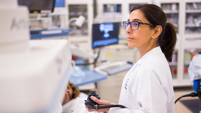
Because GIST tumors are often located in a thin layer of tissue called the submucosa, they can be missed during routine endoscopic procedures. EUS is a minimally invasive technique that relies on a thin device that uses ultrasound waves to provide clear images inside your digestive tract. EUS can be used to determine the type and stage of cancer through a tissue sample. It can help your surgeon better plan the tumor’s removal.
These diagnostic methods provide vivid, detailed images of tumors. This is critical for physicians trying to spare healthy tissue around your tumor while learning whether the cancer has spread. However, CT or EUS may not identify very small tumors or metastatic lesions (tumors that have spread from the primary tumor site).
Sophisticated Pathology Analysis
Tissue biopsies are often key to diagnosing and treating GISTs. Determining whether the cells in your tumor contain specific types of proteins, such as KIT (or CD117) and DOG1, is required to establish whether the tumor is a GIST.
Specialized physicians called cytopathologists and molecular pathologists may perform other tests to identify key characteristics of the tumor.
Our cytopathologists work together with the team performing your endoscopy or other diagnostic procedures so you’re less likely to need multiple sessions for diagnosing and treating the tumor. Tests such as next generation sequencing (NGS), which can help identify the specific molecular "on-off" switches in your tumor, will require additional time to complete.
Treatment is based on a comprehensive analysis of the GIST’s genetic mutations, location and whether it has spread to other areas of the body. Every patient’s treatment plan is customized to the specifications of their disease and overall health. Treatment for GISTs may include:
Surgery
Removing the entire tumor, either through open or minimally-invasive surgery, offers the greatest chance to be cured. We offer our patients leading-edge surgical treatments, including an array of laparoscopic and robotic surgical techniques that enhance recovery and reduce pain after surgery. Removal of lymph nodes is not necessary, but it’s important to remove the tumor intact. Rupturing the tissue that surrounds the tumor can spill tumor cells into surrounding areas, which can then grow uncontrolled. That’s why it’s critical to see experienced GIST surgeons like those at the Center for Gastrointestinal Oncology.
Non-Surgical Treatment
Non-surgical treatments involve either imatinib mesylate, sunitinib, regorafenib or novel agents being tested in clinical trials. Imatinib has dramatically improved treatment outcomes for adults with GISTs — a targeted therapy directed at the principal molecular abnormality driving the tumor. Unlike traditional chemotherapy, which destroys both cancerous and non-cancerous cells, imatinib controls GISTs for an extended period in many patients. It has minimal side effects and can be used before surgery to shrink the tumor. After surgery, it can be used to help prevent the cancer from returning. Patients unable to have surgery will generally be given imatinib. After months or years of imatinib, GISTs may become resistant and other drug regimens may be recommended. We routinely use next generation sequencing to identify the specific molecular drivers in an individual patient’s GIST. This can help us inform you of the likelihood your tumor will respond to therapy and the optimal dosage for the drug.
Life Raft Group: GIST support group dedicated to finding a cure through research and patient support.
GIST Support International: Non-profit organization that provides support as well as educational and financial resources to GIST patients.
Convenient Locations for Cancer Care

Cancer Care Second Opinions
Request a second opinion from UChicago Medicine experts in cancer care.

Why Choose Us for Cancer Surgery?
Our surgeons offer sophisticated techniques, including minimally invasive and robotic options, to treat cancer.

