Interventional Pulmonology & Advanced Bronchoscopy
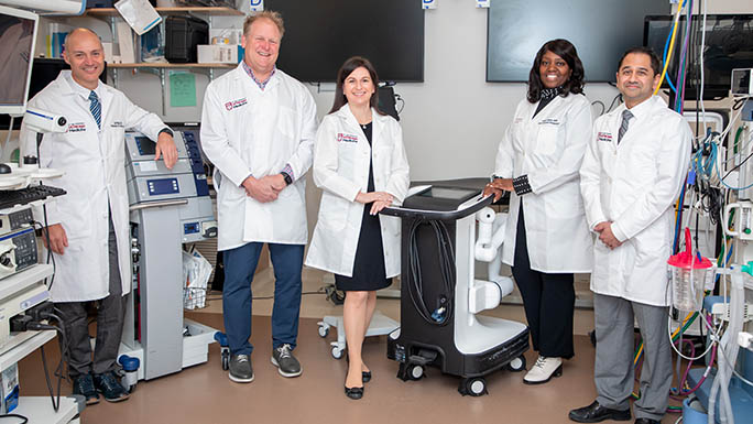
Many lung diseases can be diagnosed and treated without surgery using advanced bronchoscopy. During these outpatient procedures, interventional pulmonologists pass a thin tube with a tiny camera (or scope) through the nose or mouth and into the lungs.
The University of Chicago Medicine is a leader in the field of bronchoscopy. Our interventional pulmonologists have decades of experience and are highly committed to improving care for people with lung problems. They continuously research new ways to treat patients by regularly engaging in clinical trials to test advanced approaches and emerging technologies.
As a result, our pulmonologists have repeatedly been the first in the Midwest to adopt state-of-the-art bronchoscopy methods. For instance, they have more experience than any other Illinois hospital in using robotic bronchoscopy, a breakthrough approach that allows pulmonologists to reach areas deep within the lung that were previously only accessible with surgery.
Our patients also benefit from timely and compassionate care. For example:
- UChicago Medicine provides bronchoscopy procedures five days a week (Monday through Friday). As a result, we can schedule patients for their procedures in less than a week, whereas most medical centers in the Chicago area schedule about a month out.
- Our team provides comprehensive services before, during and after a patient’s bronchoscopy. Our physicians and nurse practitioner educate patients and family on what to expect during and after the procedure. We also arrange any follow-up care or supplies that may be needed at home.
- Finally, we collaborate with the patients’ other physicians and caregivers to ensure that all care is coordinated. This team approach helps ensure that our patients benefit from the combined expertise of a variety of medical experts.
Diagnosis. Our interventional pulmonologists use sophisticated robotic bronchoscopy to determine whether a lung nodule (abnormal growth) is cancerous. Using a 3D roadmap of the patient’s lung, which is developed from imaging scans, our pulmonologists guide a flexible endoscope equipped with a camera and small tools through the patient’s mouth to the nodule in the lung. The pulmonologist then biopsies the nodule, or collects a sample of tissue, which is immediately lab tested for cancerous cells.
Staging. Our physicians are also leaders in using minimally invasive methods to biopsy lymph nodes in the chest cavity and lung — a key step in the staging of suspected lung cancer.
The process, called transbronchial needle aspiration, may be recommended after a CT/CAT scan reveals an abnormal lymph node, which may indicate lung cancer. Conventional lung biopsy methods usually require surgery to reach a lymph node and extract a tissue sample for biopsy testing.
With transbronchial needle aspiration, there is no need for exploratory surgery to reach the lymph node. Instead, the physician can pass a special needle through the bronchoscope to reach the specific lymph node and extract tissue for biopsy. This advancement means patients can avoid unnecessary diagnostic surgery.
Transbronchial needle aspiration is done on an outpatient basis and usually takes less than an hour to complete. Patients return home later the same day.
Management of cancer complications. When patients with lung cancer have trouble breathing, our pulmonologists can provide relief, using advanced bronchoscopy procedures to open up the airway. We offer tumor debulking by laser and cooling methods. We also offer a unique treatment using photodynamic therapy (PDT) to accurately destroy tumors that are encroaching into airways. Closed off structures can be dilated, and then a stent is placed inside them, helping patients breathe easier.
UChicago Medicine is recognized as one of the top medical centers in the United States for placing valves in the lung using BLVR. Our physicians were involved in prior research trials that helped bring these technologies to the United States.
Our interventional pulmonologists are experts in performing bronchial thermoplasty, which is now approved by the FDA. The procedure involves applying heat to the airway and lungs to smooth muscle tissue. This helps minimize airway constriction during asthma attacks.
Our interventional pulmonologists also use advanced bronchoscopy approaches to diagnose, treat, and relieve a variety of noncancerous lung and airway problems, including:
- Sarcoidosis
- Pleural disease or pleural effusion
- Interstitial lung diseases
- Lymphadenopathy (swollen lymph nodes)
- Infections within the lungs
- Complex airway diseases, such as bronchial stenosis and tracheobronchomalacia
Interventional Pulmonology Team
Sampling and Evaluating Lung Nodules and Masses: Expert Q&A
Pulmonologists D. Kyle Hogarth, MD, and Ajay Wagh, MD, talk about different ways physicians can detect and diagnose lung nodules and masses, including advanced bronchoscopy techniques that do not require incisions or surgery.
[MUSIC PLAYING] Hello, and welcome to At The Forefront Live. A lung mass can be a frightening discovery. However, not everyone who receives an abnormal CT scan should be rushed into surgery. Today there are better insights into cancer and other lung diseases. Advanced technology and minimally invasive options are available.
Dr. Ajay Wagh and Dr. Kyle Hogarth will discuss the latest in lung nodule diagnostics, management, and treatment. And as always, we'll take your questions during our 30 minute program. That's coming up right now on At The Forefront Live.
[MUSIC PLAYING]
And we want to remind our viewers that today's program is not designed to take the place of a visit with your physician. Let's have each of you start off by introducing yourselves to our audience, and tell us a little bit about what you do here at UChicago Medicine. And Dr. Hogarth, we'll start with you. It sounds like you're in a busy, busy place.
I apologize.
It's OK.
I'm actually in the endoscopy suites. So my name is Kyle Hogarth. I am a Professor of Medicine here. And I have been working at the University of Chicago since 1998. And was fortunate enough to start the bronchoscopy program here, and the Nodule program. And basically work very hard to make sure that patients get the answers that they need to help decide what's the best next path when they find an abnormal CT scan.
Because an abnormal CT scan is terrifying. And you want to have something reliable in what to do next.
See, this just shows how important it is that we do these programs here. Our doctors will actually even join us from the places where they're doing the work. Dr. Wagh, let's hear a little bit about you. We just talked a moment ago, and you're pretty new here.
Yes, sir. Well, my name is Ajay Wagh. I'm new here to the University of Chicago, and very thankful to be here. I'm grateful to participate in Dr. Hogarth and Dr. Perdue's team. We have a great team here, and I'm excited to be part of it.
I recently completed an interventional pulmonary fellowship, which brought me here. And prior to that, I was a private practice pulmonary critical care doctor for six years. So I'm excited to be here in the city, and part of this program.
Well, we're very happy to have you. And of course, you came here at kind of an odd time, during a pandemic. But I'm sure you'll enjoy UChicago Medicine. It's a wonderful, wonderful place. And the city of Chicago is a great place and a lot of fun.
So let's start off with our questions. And Dr. Hogarth, I want to start with you. And if you can, just kind of set the stage for us and tell us a little bit about nodules and masses, and what are they and how do people even know that they have such a thing in their lungs.
Yes, sir. So if you have an x-ray or a CAT scan, there is a chance that the word module or mass is going to show up in the report. And it's something solid. If you think about it, the lung is mostly air. And so think of it like a sponge. So something solid inside the lung needs an explanation, because there shouldn't be something solid in the lung.
But there's many things it could be. And without a doubt, the possibility of cancer is what scares everybody. But the first thing to understand is there's a long list of not cancer reasons you could have a nodule in your lung. And teasing out what's what is what Ajay and I do.
He and I, and our other partner, Dr. Mergue, work very hard to make sure that if you need a procedure, it's the right procedure. If you don't need a procedure-- because there's no chance that this is cancer-- we would like to avoid doing anything invasive on you. And I think that's the first key step.
Getting an expert opinion about what could this nodule actually be. And using some of the tools that we have. And then if we do need to do a biopsy, making sure the correct biopsy gets done. So that you get an answer as to what this nodule actually is.
You know, and I want to talk a little bit more about biopsies here in just a minute. Because it's interesting how you do them in the lung. We're going to get to a little bit more detail of that one here in just a moment. But Dr. Wagh, can you talk to us a little bit about just-- I think as Dr. Hogarth just mentioned, if somebody comes in and sees a physician. And they hear, oh my gosh, I've got a nodule. The immediate reaction is you're probably frightened.
Is that-- should you be frightened? Or is this something that happens and you just need to get it checked out? Or is that the moment of panic at that point?
Well, I think that there's several possibilities. And I think we like to take things one step at a time. Panicking, obviously, is never helpful. And I think what we want to do is offer a pathway here in our program for patients to get everything they need. And we will kind of shepherd the patient along the way. And that's kind of comforting, I think, for most patients.
So I think first step is don't panic. And then second step is find the right people to help take care of you. And we're, of course, happy and eager to help. And I would say the only other thing, as a pulmonologist, is if you smoke, try to stop. And we can help you do that, too.
You know, and I was only being partially facetious when I said panic is kind of the natural-- because I think for a lot of people that's just the natural reaction.
Absolutely.
And you don't want to. You want to be calm and cool. And you two, and your teams, are really good at helping people through that situation. Because initially when you're faced with something like that, everything kind of just goes over your head. But that's part of what you do. And Dr. Hogarth, I don't know if you can talk to us a little bit about how do you work with the patients?
Because I know this is a very complex situation. Communication is important with the patients. Can you kind of walk us through that? What happens?
Sure. You know what, I always tell people is there is a long list of things that the nodule could be. But in reality, if you're a patient, there's only two things. It's either cancer or everything else. And so part of our discussion is, what's the probability that this nodule that you have on your CAT scan-- is it actually cancer or not?
What's that chance? And we have a series of other tests we can do. Some of them are blood based tests. Some of them are just re-evaluating the CAT scan you have. There are characteristics of nodules that make them more concerning for cancer, as opposed to less concerning.
So first is just a discussion with you of what is the probability that this could be a malignancy for you. And this is important. Because in some cases, our plan for you is to get a follow-up CAT scan, is to do watch and wait. And you say, well, wait. It could be cancer. The probability, if it's low enough, we don't want to do invasive things to you. Because the chance it's cancer is so low, and every invasive procedure always carries a risk.
So if the risk of cancer is low, but the risk of a complication is the same, I don't want to harm you, right? First, do no harm. It's an oath both of us took.
So we do want to remind our viewers, we'll take your questions for our experts. Just type them in the comments section. We'll try to get to as many as we can over the next half hour. Dr. Wagh, you mentioned as a pulmonologist you tell people stop smoking. That's going to be number one on the list. And I would imagine in this-- I've got to word COVID of in here, because you know, it's what we're talking about everywhere.
That's right.
That's another thing that you probably want to caution people about. Stopping smoking can help you just across the board. And that would be another area, I would imagine.
Yes, sir. I mean, I think we are living in a strange time. And smoking is certainly a problem, a historical problem that we're working to deal with every day. And you know, COVID makes it harder for patients to see doctors. But we do have avenues to help with that. I mean, we do have telemedicine options. And there are potential treatments to help patients quit smoking as well.
But you know, I think that there's so many different possibilities when it comes to management that we're quickly learning how to utilize technology, even in telehealth, to help patients get what they need.
Yeah, and I want to tell people-- this is a very, very safe place. We're still operating. We're open for business. And probably the worst thing that could happen is that somebody would forego treatment that they need because they're afraid of COVID. We don't want that to happen. So--
Oh, let me reinforce that.
Absolutely, yeah.
The hospital is safe, the hospital is clean. I work here, I go home, I kiss my children. I kiss my spouse. Like, I'm not worried about spreading disease. We are extremely cautious about everything here. And we are going to be first and foremost interested in protecting you, as well as protecting ourselves and our staff.
But to delay any amount of care. You know, you said at the very beginning, I have a nodule, should I panic? No, don't panic. But also don't ignore it, and don't delay it. And as Dr. Wagh just said, we are able to do video visits and televisits. I can meet with you virtually. If it bothers you to come near the Medical Center, fine, let's do it via the computer. And let's go through your CAT scan and let's have this discussion about what our next step is.
You know, in fact, just to even further hammer home that point. Even the show that we're doing right now, you two are remote. We're in very separate areas. I'm in the studio all by myself, as you can see here. There's nobody else here. We don't even have any camera people in here. That's why I'm not moving a lot, not that I move a lot anyway.
But we're very careful about that. And we're very serious about that. And it's important here. We are taking questions from viewers. We do have one that I want to get to. And either one of you can jump on this one. Maybe Dr. Hogarth, you can start. The question is, how quickly do cancerous lung nodules grow compared to other types of cancer? And how urgently must patients act?
No, it's a great question. The fear always is that cancers are going to grow. Every tumor, of course, has its own biology speed at which it grows. But generally speaking, a lung cancer-- when someone says to you, hey, we want to get a follow-up CAT scan, the reason they're suggesting that is that the nodule you have is so small or has characteristics that are so convincing that it's benign, that that two or three month interval that they've suggested-- if I'm wrong and it's actually a cancer, the amount that it's going to grow in that time period is so small that we've not lost anything.
You will still be the same stage. And what we've gained, of course, is for all of those scans, that nothing changed and you avoided an unnecessary invasive procedure.
And Dr. Wagh, maybe you can take this next one. What are some of the options to evaluate lung nodules and lung masses? We can talk about imaging modalities. And Dr. Hogarth mentioned blood tests even, a few moments ago.
Yeah, there's several possibilities in that regard to evaluate these. I mean, the first thing is first, is we do have lung cancer screening, which we offer patients CAT scans if they're eligible and have a smoking history. And you can speak with your physician about that. Or come and visit a lung physician. But many times, you might notice something on an x-ray that's not part of the screening pathway. But a doctor may see something on a chest x-ray.
But also cat scanning. And then there are other types of imaging techniques, like PET scans, other images that we use to evaluate lung nodules. And so those are our mainstays of imaging. But there's many other tests. Dr. Hogarth kind of briefly said something about the blood tests. But of course, there's biopsies.
There's all kinds of different tests. We even use-- in order to evaluate a patient's risk-- we use calculators to help evaluate that too based on a patient's history and imaging findings.
So-- go ahead, Dr. Hogarth, did you have something you wanted in?
Just to echo what Dr. Wagh said. There's large databases that have been built off of the experience of radiology to be able to essentially plug-in and give a number. Hey, this nodule has a 20% chance of being cancer, which is not a number anybody wants to hear. But of course, there's an 80% chance it's not cancer.
So before we go and suddenly just remove a whole portion of your lung, let's slow down for a second and do things right. Some of the blood tests we have, have the ability to change that number. You know, we go, oh, it's a 20% chance. Well, the blood test actually showed that it's less than 5%. Oh, less than 5%, OK, let's slow down a little bit.
The whole key thing, too, is that this is an ongoing dialogue between us and the patient. We're not going to just say, you must do this. We're going to give you some strong recommendations. But we're also going to work with you. And every patient is different. Maybe a 3% chance of cancer is acceptable to some, and terrifying to others, and everywhere in between. But we also want to explain to you what we're going to do to actively follow you.
What you're never going to hear from us is to say, now there's nothing to do, leave. We're going to tell you a outlined plan that is backed up with data as to why we're doing this.
So I want to get back to biopsies for just a moment. And we had a question from a viewer that dovetails perfectly into what I'm kind of curious about. And that is how biopsies work. And Janet wants to know how invasive is a lung biopsy? And I was fortunate enough, I think, gosh, it's been over a year ago. It was way pre-COVID, but you showed me one piece of equipment that you had, Dr. Hogarth.
It was pretty fascinating to see what you could do inside of a person's lung with a very, very minor, very minor invasive procedure. I mean, it's really amazing. Can you kind of talk to us a little bit about that, and walk us through that?
Yeah, sure. And then I'll have Ajay go at it as well. So look, there's three ways to sample inside the lung. There's a surgeon, who's going to go in and cut part of it out. And obviously, you know, even with minimally invasive surgery, it's still a surgery. They're still cutting in you. There's also what's called a needle biopsy. Where it's basically put right through your chest into the lung nodule done through the radiology department.
And these procedures all have their own benefits, but also their own complications. What Dr. Wagh and I do is a procedure called bronchoscopy. So we go through your mouth. Your lungs are going to be ultimately attached to your mouth. Why aren't we just following the pathway down? We're fortunate enough here at UChicago Medicine to have a robotic endoscope that lets us get to parts of the lung we've never been able to get to before.
We use that CAT scan, build a three dimensional map of your lungs, and we drive to the spot where that's at. Pass instruments out, take little pieces that we-- so you're not going to miss anything, you know, volume wise. And we do it through your mouth. So there's no cutting. So when we're done, you go home. And our complication rate is the lowest amongst the three. And we have a high success rate to get you an answer.
And where this matters is, of course, if I go and prove that it's not a cancer, then rather than being cut open and proved it was not a cancer-- which is great it's not cancer, but you've been cut open. Instead, you might have a little sore throat for a day or two. That's not hard to convince someone.
So Dr. Wagh, it was interesting because this is almost like a video game. I remember when Dr. Hogarth showed this to me. It's almost, you know, again it's like a video game, science fiction, it's pretty amazing. Can you talk to us a little bit about what the patient experiences in this procedure? And how minimal it actually is?
Yes, so a patient typically comes in basically just for a few hours during the day. You can't eat after midnight. But you come in, we have a pre-procedural area where the patients get kind of their IV. And sign a few papers. And then they come to our lab. And we are lucky enough to have anesthesiologists who help take care of the patient during the procedure.
And then we go in with our scopes. And we get the tissue that we need. And the patient goes afterwards to a post-procedural area, where they recover. And hopefully, go home if nothing happens. I do think that it's worth saying that complications are pretty rare with the scopes, the endoscopy that we perform.
And I do also think it's worth mentioning that by doing the bronchoscopy, as opposed to choosing an alternative technique, such as a needle biopsy, we're also able to evaluate the lymph nodes in the chest. And that's a very important part for a cancer evaluation. And so that becomes one procedure, as opposed to multiple procedures.
So talk to us a little bit more about the lymph nodes. And either one of you can do that. What exactly goes on there, and why is that so critical?
Well, if you have a cancer, the next question is, what stage is it? Right? And so the lymph nodes are where cancer would spread to first. So if we think you're at early stage cancer, that's great. I want to know you're an early stage cancer. And so as Dr. Wagh just pointed out, in the same procedure, after we've just proved that is a cancer, we're going to then go sample your lymph nodes.
So we'll wake you up. I'm not happy that I have to tell you it's cancer. But what I can also tell you is it's cancer, here's what stage it is. And so now you're going to go to the surgeon to be cured. Or you're going to go to radiation or whatever. You don't have to go get another procedure that's going to take time to then figure out what stage you are.
I'll also point out that our procedures, like Dr. Wagh said, are done under anesthesia. This isn't that twilight. You're out. You will not know we're doing this to you. You are comfortable. We're going to do our work. You're going to go home.
So I have two from viewers that I have to pass along. These are not questions. But I love these. So I always have to do this. Elizabeth says, I've had several bronchoscopes done by Dr. Hogarth, he's amazing. So that's nice. Karen says, your pulmonary department is the best. So a little bit of a fan club going here, but that's awesome. Now, a question.
We'll get you a speech card.
[LAUGHTER]
We could get you a plaque or something. Well, that's nice. And again, in all seriousness, I think that shows really, you know, the work that you do with the patients. And that's very important. Because it's a difficult time in people's lives when they have something like this done. So this is an actual question.
Amit, I hope I'm pronouncing this correctly. Can an 11 millimeter nodule be biopsied by that bronchoscope method through the throat?
Go ahead, Ajay.
Well, it certainly can. When we-- and I'll also say it depends. But we can. 11 millimeters is rather small. And that's sort of when we take a look at the CAT scan very closely. We evaluate whether or not it's a target that we can reach. We look at the airways and other parts of our computer modeling to see if that's something that we can get to.
And we also try to figure out, is it a lesion that requires biopsy? Or should we offer something else? If you're concerned about cancer and there's an intermediate pre-test probability, based on a calculated evaluation, then we can potentially offer a blood test or something else that may potentially reduce the risk. Or suggest that the pre-test probability is lower. So--
Interesting. Another question from a viewer, and this is Carla. And this is a little bit inside baseball. So I'm going to have you answer the question, but also kind of explain what she's asking here. Is following a nodule ground glass opacity with yearly CT standard? When there are no changes from scan to scan. And I don't know. Dr. Hogarth, do you want to start on that one?
Sure, so a ground glass nodule is definitely a different thing than a very solid nodule. A ground glass nodule almost looks like some wispy smoke on the CAT scan, if you will. So ground glass nodules are a different biology. In some cases, they are a precancerous lesion. In other cases, they are actually a cancer.
But for many people are extremely, extremely slow growing cancer. And one that has a very low invasive potential. Meaning, it's technically a cancer, but it's never going to necessarily bother you. And one of the reasons we do this yearly image, because it is so slow growing, if it's not changing year after year, then the probability that it's going to do anything to you becomes so low that we actually leave you alone.
Because why would I put you-- why would I cure you of something that's never going to harm you? Now, these are complicated discussions. You know, it's not just like, yeah, you do this. But when it's time to get a follow-up scan, the reason, ultimately, for these ground glasses, why they settle into yearly, is precisely because they're slow growing.
If we keep scanning you, we're never going to see change. And we're also going to just keep radiating you. Now, solid nodules, depending on the size, there are guidelines that suggest the interval of scans. And we keep spacing that interval of scan out if nothing has changed. Obviously, if things change, then that's a discussion towards biopsy.
That ground glass, if it gets larger or denser, then it's changing. And the individual tumor biology is changing. So we need to get going and do something about it.
Interesting. So Dr. Wagh, you touched on this a little bit before. But can you kind of walk us through what people can expect before, during, and after one of these procedures.
Yes, sir. So typically we'll have a clinic evaluation. And that could be in person. Or it could be a telemedicine visit. And then based on that discussion, we would set a patient up for a procedure. And we kind of-- we have a nice staff who will kind of walk the patient through what they should expect. And usually we discuss medications, if the patient is on a blood thinner. Whether they, you know, the fact that they can't eat the night before because we do general anesthesia for many of our cases.
And then afterwards, once we settle on a date, the patient comes in. They come into the sky lobby here at UChicago. And then they wait to be brought to the pre-procedural area. And at that point, they'll meet the anesthesiologist, the nursing staff. And they'll double check everything. Make sure everything looks right, that it would be safe to proceed.
And then at that point, we would bring the patient back to the our laboratory. And there we perform our procedures. And then once that's completed, we send the patient to the post procedural area, where they recover for a couple hours. And then they just go home. It's usually about a half day's worth of time.
Interesting. And so Dr. Hogarth, we have another question from a viewer. This is from Therese. And it is, would my annual low dose CT lung cancer screening show nodules? Or does it have to be a higher dose CT screening?
That's a great question. No, it will show the nodules. That's why we do it. The fact it's a low dose is because you are being screened, there is no other reason we're scanning you. So we want to-- I mean, we want to do this for everybody. We want to minimize radiation. But if it shows anything of any concern, especially if it's your first one, that may require a follow-up scan in a shorter interval or one with slightly higher radiation.
I should point out, the amount of radiation you get from a CAT scan at a center like ours-- so it has everything to do with the quality of the scanners. And as you can imagine, a place like UChicago Medicine, we've got the highest quality CT scanners. We're giving you the least amount of radiation, even for what's called a diagnostic scan. Because it has everything to do with the quality of the machine for the radiation that goes through.
So you're going to get way more bang for your buck literally as a scan by coming here. But one of the other things we were talking about, the patient journey. So Dr. Wagh and I have our partner, Dr. Mergue. We also have literally the world's greatest nurse practitioner, Kimberly. And between the four of us, we're all in clinic at any given moment.
So if you have an abnormality, the other thing that sets this place apart is if you call a regular hospital and say, hey, I've got a lung nodule, can I see somebody? You will get seen three to four weeks from now. And the national standard is roughly five weeks. You will get seen within a week every time here. And if someone ever by mistake says to you, yeah, they can see you in three months. You need to raise a fit.
Because we will always see you. We will overbook you. You will never be told, you've got to wait around to be seen after someone's told you that you might have cancer. That is not acceptable to make you wait.
I can't even imagine what that would be like if you're worried that you have cancer, and then you're told you have to wait for an extended period of time. It's got to be terrible.
Exactly.
So we talked a little bit about just screening for lung cancer in general, and what people need to know, because I know there are some folks that will go through a regular process of screening. I don't know who wants to take that one. Go ahead, Ajay.
Yeah. So I mean, we do have a regular process of lung cancer screening. We want to find patients who have a history of smoking, quit within the past 15 years. Age is usually 55 to 80. And you know, those patients typically are eligible for low dose lung cancer screening. And that would be annually until they kind of exit out after that 15 years.
It is covered by insurance. And you know, it is extremely valuable. Now, the low dose lung cancer screening has its own set of guidelines that helps us to monitor and follow any suspicious nodules. So follow-up scans could also be low dose as well. And so I do think it needs to be corrected that you should not get a chest x-ray as a screening tool. It should be a CAT scan if you are eligible.
You know, you mentioned that being covered by insurance. Is the evaluation and procedure that we've been talking about, is that covered by insurance as well? That's always the question people want to know.
No, for sure. Our fancy robot that's going to let us go everywhere in the lung is definitely covered by insurance.
That's good to know. Well, gentlemen, we're out of time. You were fantastic. You shared really some good information with our audience. So appreciate that. And thank you to our viewers for your great questions. Really, really good questions today. Please remember to check out our Facebook page for our schedule of programs that are coming up in the future.
Also, if you want more information about UChicago Medicine, take a look at our website at uchicagomedicine.org. All kinds of fantastic information there. It's a wonderful website. And it also has a lot of great COVID information. So if you need an appointment, give us a call at 888-824-0200.
We want to remind people, very important, do not forego medical care during COVID. It's so important. This is a safe place. And remember, you can schedule your video visit by also going to the website. Thanks again for being with us today. And I hope you have a great week.
[MUSIC PLAYING]
Request an Appointment
We are currently experiencing a high volume of inquiries, leading to delayed response times. For faster assistance, please call 1-888-824-0200 to schedule your appointment.
If you have symptoms of an urgent nature, please call your doctor or go to the emergency room immediately.
* Indicates required field